Adenomyosis – A research review
Adenomyosis is a clinical enigma in gynecology due to its vague etiology & pathogenesis, although multiple factors and various hypotheses are proposed related to it. It is a gynaecological problem presenting with menorrhagia, pelvic pain and dysmenorrhea.
updated on:2024-08-14 15:57:31

Written by Dr. Sanjana V.B Bhms,dbrm,cdn
Founder & medical director of siahmsr wellness.in
Reviewed by SIAHMSR
All rights reserved with siahmsr digital healthcare[siahmsr wellness]
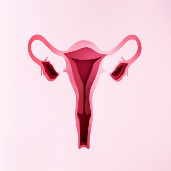
Adenomyosis
Adenomyosis is a condition affecting women of reproductive
age group, presenting with menorrhagia, pelvic pain and dysmenorrhea. It is
diagnosed by imaging studies [MRI, Ultrasound] as a benign condition
characterized by the presence of ectopic endometrial glands within the
underlying myometrium. It is found in some cases that adenomyosis and
leiomyomas coexist in women. Although
adenomyosis continue to be a serious health issue related to women’s health,
research studies related to its management is very few.
History
The origin of the term adenomyosis is from adenomyoma” in 1860 by the German pathologist Carl von Rokitansky, who found endometrial glands in the myometrium and subsequently referred to this finding as “cystosarcoma adenoids uterinum”.
Thomas Stephen
Cullen in the 19th century fully researched the 'mucosal invasion' in this
clinical condition and clearly identified the epithelial tissue invasion of
uterine mucosa.
Frankl created a
name for the mucosal invasion of the myometrium and clearly described its
anatomical picture; he called it 'adenomyosis uteri'.
The latest
definition of adenomyosis is by Bird as the benign invasion of endometrium into
the myometrium, producing a diffusely enlarged uterus which microscopically
exhibits ectopic non-neoplastic, endometrial glands and stroma surrounded by
the hypertrophic and hyperplastic myometrium.
It is commonly found that adenomyosis may coexist with pelvic endometriosis; however the significance of myometrial lesions in producing clinical symptoms, such as infertility and pain is still obscure. Furthermore, recent studies point out that adenomyosis is a progressive disease that changes in appearance during the reproductive years.
Clinical presentation & risk groups
Adenomyosis may present with menorrhagia, pelvic pain and dysmenorrhea. Dyspareunia & infertility are also symptoms of this clinical condition.
Adenomyosis may be focal or diffuse type depending on its
distribution within the myometrium. Diffuse adenomyosis is defined by the
presence of multiple foci within the uterine myometrium, while focal
adenomyosis appears as isolated nodules of hypertrophic myometrium and ectopic
endometrium.
A large number of
cases of adenomyosis are found in multiparous women as pregnancy may lead to
the formation of adenomyosis by allowing adenomyotic foci to be included in the
myometrium due to the invasive nature of the trophoblast on the extension of
the myometrial fibers.
Furthermore, adenomyotic tissue may have a higher ratio of
estrogen receptors and pregnancy may facilitate the development of islands of
ectopic endometrium.
An enhanced risk
associated with prior uterine surgery in women with adenomyosis is
inconsistent. According to some studies adenomyosis results when endometrial
glands invade the myometrial layer, with surgical disruptions of the
endometrial-myometrial border.
Another study [6]
shows that dilation and curettage procedures in gynecology are associated with
higher rates of adenomyosis than women without pregnancy terminations.
There is a
controversial finding [7] that adenomyosis prevalence is low in smoking women.
Probably the reason may be decreased serum levels of estrogen in smokers, and
adenomyosis has been suggested to be an estrogen-dependent disorder. However,
there is also evidence that there is no association between adenomyosis and
smoking.
Adenomyosis is not
common in postmenopausal women but a higher incidence of adenomyosis has been
reported in women treated with drug tamoxifen for breast cancer. It is
suggested that the prolonged and unopposed estrogen-like stimulation by
tamoxifen may play a causal role in the development of adenomyosis [10].
Adenomyosis may be
a risk factor for the development of intramural ectopic pregnancy [8].
The presenting symptoms of adenomyosis are non-specific
often and these can also be found to be associated with disorders such as
dysfunctional uterine bleeding, leiomyomas and endometriosis, among others.
Adenomyosis and leiomyomas commonly coexist in the same woman [15 and 57 %].
Mostly the preoperative differentiation of both conditions
in the same uterus is difficult, even with the addition of imaging techniques
including ultrasound and magnetic resonance imaging. However post hysterectomy
studies show that women with adenomyosis have been shown to have lower uterine
weights, more dysmenorrhea, dyspareunia, pelvic pain and more disease-specific
symptoms compared to women with leiomyomas alone. Therefore clinicians should
keep in mind the differential diagnosis of adenomyosis in women with symptoms
that seem disproportionate to the level of leiomyoma disease alone.
Diagnostic accuracy and technological
aid in adenomyosis
Ultrasound [TVS]
and MRI imaging studies give clear cut data about adenomyosis. According to
imaging studies the junctional zone myometrium can be clearly distinguished
from the endometrium and outer myometrium, and diffuse or focal thickening of
this zone is now recognized as one hallmark of adenomyosis.
Of late magnetic
resonance imaging provides superior soft tissue resolution and considered as
the most accurate technique for non-invasive diagnosis.
Adenomyosis
represents a spectrum of lesions, ranging from increased thickness of the
junctional zone to overt adenomyosis and adenomyomas, which in turn can be
subclassified.
Bird et al. first classified adenomyotic lesions according
to the depth of penetration on uterine myometrium.
Grade I represent adenomyosis sub-basalis/sub-endometrial
basalis adenomyosis within one low-power field below the basal endometrium, but
with no further penetration.
Grade II represents adenomyosis penetration to the
mid-myometrium.
Grade III represents
adenomyosis penetration beyond the mid-myometrium.
In addition to
using the percentage of myometrial penetration, Sammour et al. classified the
degree of spread of adenomyosis by the number of foci and the extent of
disease. They also found a direct correlation between the spread of adenomyosis
and dysmenorrhea, but not the depth of penetration.
Adenomyosis in adolescents & young women
Recent studies
based on MRI criteria for diagnosis suggest that the disease may cause
dysmenorrhea and chronic pelvic pain in adolescents and younger women of
reproductive age.
Cystic adenomyosis is a rare cause of abdominopelvic pain
and dysmenorrhea in adolescents. Imaging study is important in distinguishing
this disease from other gynecological disorders.
Management
Medications
Some hormonal pills that are used include non-steroidal
anti-inflammatory drugs and/or hormonal therapy [oral contraceptive pills,
high-dose progestins, a levonorgestrel-releasing intrauterine device, danazol,
gonadotropin-releasing hormone agonists] etc. to manage adenomyosis in young
women.
Adenomyosis is commonly managed with hysterectomy in
perimenopausal and postmenopausal women as the minimally invasive surgical
techniques such as endometrial ablation/resection, myometrial
excision/reduction, myometrial electrocoagulation, uterine artery ligation etc.
have limited success in the treatment of adenomyosis.
Hysterectomy is the treatment option in cases failing to
control dysmenorrhoea, menorrhagia and dyspareunia with medications and
hormonal therapy.
Recently new techniques including uterine artery embolization (UAE) and magnetic resonance imaging guided focused ultrasound (MRgFUS) show promise in treating adenomyosis.
Complementary & alternative
medicine
Homeopathic medicinal management
Apis mellifica,
caulophyllum, Ferrum met , Lachesis, , pulsatilla, platina, thuja, Sabina,
Sepia are the medications frequently used to manage the symptoms and
complications of adenomyosis.
Medicines are chosen based on individualization and symptoms and signs manifested by the disease. A few medications help to control menorrhagea , dysmenorrhoea while others address anaemia and weakness from uterine haemorrhage. Medicines are prescribed for managing infertility and dyspareunia also.
References
1. Benagiano G, Brosens I. History of
adenomyosis. Best Pract Res Clin Obstet Gynaecol. 2006;20:449–463. https://pubmed.ncbi.nlm.nih.gov/16515887/
4. https://pubmed.ncbi.nlm.nih.gov/19539194/
8. Lu H F, Sheu B C, Shih J C. et al.
Intramural ectopic pregnancy. Sonographic picture and its relation with
adenomyosis. Acta Obstet Gynecol Scand. 1997;76:886–889. https://pubmed.ncbi.nlm.nih.gov/9351419/

Recommended For You
Adenomyosis – A research review
Adenomyosis is a clinical enigma in gynecology due to its vague etiology & pathogenesis, although multiple factors and various hypotheses are proposed related to it. It is a gynaecological problem presenting with menorrhagia, pelvic pain and dysmenorrhea.
Ankylosing spondylitis- A clinical study of complications & management protocols
Ankylosing spondylitis [AS] is a chronic inflammatory autoimmune disease that mainly affects spine joints and over the time involving other joints of the body also. AS is characterized by the involvement of the spine and sacroiliac (SI) joints and peripheral joints, digits, and entheses.
Related Links

sdfgh