Vertebral artery Stenosis - A comprehensive overview
Stenosis or occlusion of the vertebral artery can occur unilaterally or bilaterally. The arterial occlusion may cause decreased artery perfusion and as a result symptoms of transient ischemic attack, such as vertigo, ataxia, diplopia, disturbance of speech, and bilateral hemianopia may occur
updated on:2024-02-05 00:57:14
Vertebral artery stenosis – A Comprehensive
overview
Vertebral artery
The vertebral artery is typically the first major branch of
the subclavian artery on both the left and right sides of the body.
The vertebral arteries are major arteries of the neck.
Typically, the vertebral arteries originate from the subclavian arteries. Each
vessel courses superiorly along each side of the neck, merging within the skull
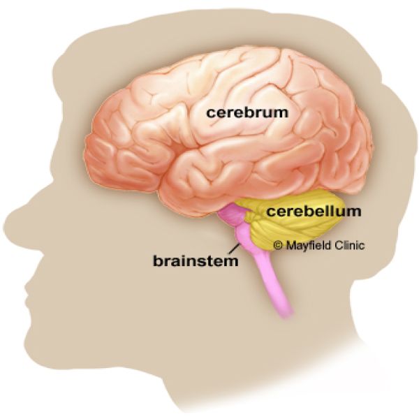
to form the single, midline basilar artery. As the supplying component of the
vertebrobasilar vascular system, the vertebral arteries supply blood to the
upper spinal cord, brainstem, cerebellum, and posterior part of brain.
Triangle of the vertebral artery is a region within the
root of the neck and has following boundaries:
· Medial border of
anterior scalene muscle (lateral)
· Lateral border of
longus colli muscle (medial)
· Carotid tubercle
(apex)
· First part of
subclavian artery (base)
The vertebral artery runs from base to apex (prior to
entering the transverse foramen of 6th cervical vertebra).
It can be divided into four anatomically different segments
(V1-V4), where segments V1-V3 are classified as the extracranial vertebral
artery, and segment V4 is considered the intracranial vertebral artery
Once the artery pierces the dura mater, it becomes the
intracranial vertebral artery. The fourth segment (V4), known as the
intracranial segment, is defined from where the artery pierces the dura mater
at the foramen magnum to where it joins with the contralateral vertebral artery
to form the basilar artery.
Vertebral artery stenosis
Stenosis or
occlusion of the vertebral artery can occur unilaterally or bilaterally. The
arterial occlusion may cause decreased artery perfusion and as a result
symptoms of transient ischemic attack, such as vertigo, ataxia, diplopia,
disturbance of speech, and bilateral hemianopia may occur.
The occlusion of
artery may also cause decreased perfusion of basilar artery and it may be
manifested with vertigo, dizziness,
diplopia, ataxia, dysarthria, nausea, nystagmus, drop attacks, loss of
consciousness, numbness, and an
increased risk of strokes or transient ischemic attacks.
These symptoms are
more prominent when the arterial occlusion is bilateral. Neck pain or headache
is an important warning symptom of VAD, and the presence of a concomitant
sensory level or Brown-Séquard syndrome is helpful for the early diagnosis of
Spinal cord infarction[SCI] caused by VAD.
Vertebral artery stenosis can also cause recurring syncope, headaches, recurrent
stroke, palsy of cranial nerves, altered consciousness, altered function of the
sensory and pyramidal tracts, cerebellar infarcts, and tinnitus.
The proximal
vertebral artery is the most prevalent location of vertebral artery stenosis
[1].
Around 20% of posterior circulation ischemic strokes
involve the stenosis of the vertebral artery [2].
If symptomatic VAS is left undiagnosed and unmanaged, it
may result in strokes, myocardial infarctions, vertebrobasilar insufficiency
(VBI), and sudden death[3].
Risk factors
· Diabetes
mellitus, hypertension, and hyperlipidemia are common risk factors for
vertebral artery stenosis.
· Cigarette
smoking
· Old
age
· Causes
of arterial occlusion
· Calcification
of arteries
· Calcification of blood vessels plays a critical role in up to 90% of atherosclerotic lesions. Calcification occurs when calcium and phosphate get deposited in the blood vessels.
· Blood
vessels are composed of three layers. The innermost layer is the tunica intima,
the middle layer is called tunica media, and the outermost layer is called the
tunica adventitia. The intimal
calcification results in atherosclerosis and stenosis.
· The
intimal calcification of the vertebral artery can cause vertebral artery
stenosis through atherosclerotic lesions.
· Atherosclerotic
Lesions
· Atherosclerosis
is the most important cause of vertebral artery stenosis. High cholesterol in
the blood triggers atherosclerosis.
· Atherosclerotic
plaques cause lesions and the stenosis of vessels
· Dissection
lesions
· Dissection
lesions are one of the primary causes of cerebrovascular accidents in
populations under 45 years old, with an approximate incidence of 10%-25%.
Vertebral artery dissections are traumatic, associated with pre-existing
arteriopathies, or spontaneous [4].
· Spontaneous
dissections are associated with old age, whereas traumatic dissections are seen
in young people. Chiropractic manipulation, spontaneous head movements,
cervical trauma, oral contraception, and fibromuscular syndromes like
Ehlers-Danlos syndrome type IV and osteogenesis imperfecta type I are the
causes leading to dissection.
· The
process of dissections happens when the tunica intima tears, blood accumulates,
and dissects in the arterial wall. As a result of the dissection, thrombosis
and hematomas precipitate. The hematomas and thrombosis can result in
hypoperfusion, stenosis, thromboembolism, Wallenberg syndrome, and strokes.
· Fibromuscular
Dysplasia
· It is
a hereditary vascular disease that is non-atherosclerotic and non-inflammatory
and causes stenosis, thromboembolism, dissections, fistulas, hypoperfusion of
cerebral blood flow, and aneurysms in any network of blood vessels.
· Giant
cell arteritis
· GCA is
an autoimmune disorder that affects the vasculature by causing inflammation at
the internal elastic membrane.
· Bony Compression
Vertebral arteries may become
stenosed or occluded due to being compressed at any point along the course of
the vertebral artery [5].
· The
most common anatomical point prone to extracranial compression is at the C1-C2
level due to its association with the atlantoaxial joint and membrane.
· Patients with atherosclerotic arterial disease and asymptomatic vertebral artery stenosis have a higher risk of posterior circulation ischemic stroke than patients without such a stenosis, but the absolute risk remains low.
Clinical presentation
·
Asymptomatic stenosis of the vertebral artery.
There is a low risk of posterior circulation stroke in
patients with asymptomatic vertebral artery stenosis.
· Subclavian
steal syndrome (SSS)
It is the occlusion of the subclavian artery, closest to
the origin of the vertebral artery, results in the reversal of blood flow in
the ipsilateral vertebral artery and shunting of blood from the contralateral
vertebral artery into the subclavian artery.
· Vertebrobasilar insufficiency (VBI)
VBI occurs due to hypoperfusion of the vertebrobasilar
arterial system resulting in vertigo, diplopia, dizziness, loss of
consciousness, dysphagia, dysarthria, nausea, ataxia, nystagmus, and numbness
[6].
Vertebral artery stenosis is also associated with a high
risk of strokes, myocardial infarctions, and sudden death [7].
The symptomatic presentation of vertebral artery stenosis can vary and can present with vertigo, diplopia, dizziness, nystagmus, nausea, ataxia, and numbness [6]. If vertebral artery stenosis is left undiagnosed and untreated, there is a high risk of cerebrovascular accidents, myocardial infarctions, and sudden death.
Investigations
· Duplex
ultrasonography (DUS) is the most common and standardized test for
the initial screening and diagnosis of vertebral artery stenosis (VAS) and
subclavian steal syndrome (SSS) as it is a safe, accurate, non-invasive, and
cost-effective diagnostic method
· Computed
tomography angiography (CTA) is a non-invasive imaging
tool to evaluate vertebral artery stenosis and disease [12]. CTA is superior to
DSA because it can image the extracranial portion of the vertebral artery while
avoiding the potential complications of using catheter angiography.
CTA is not the best imaging technique for patients who have a history of renal disease due to the high proton intensity and radiation that it exposes patients to.
·
Magnetic resonance angiography (MRA)
It is similar to CTA, and is also a
non-invasive imaging technique used to evaluate vertebral artery stenosis and
disease [12]. Imaging severe stenosis of the vertebral artery, such as the
ostial portion, poses a challenge for MRA. MRA overestimates the stenosis of
the ostial vertebral artery; however, the use of contrast mediums resolves this
challenge. Contrast-enhanced MRAs (CE-MRAs), compared to catheter angiography,
yield higher-resolution images of all extracranial cervical vessels .CE-MRA
also has higher sensitivities and specificities for stenosis of the
extracranial vertebral artery than MRA without contrast or DUS. However, CE-MRA
cannot be used with patients who have pacemakers or other metallic devices.
Management
The chronic management by modern or conventional medicine include:
· Antihypertensive treatment is recommended to treat hypertension by lowering blood pressure below 140/90 mmHg.
· Statins with or without LDL-lowering therapy and bile acid sequestrants or niacin are used to treat hyperlipidemia by lowering LDL cholesterol below 100 mg/dL.
· Physical activity, glucose-lowering medications, dieting, and a statin-type lipid-lowering medication are prescribed.
· It is recommended that patients diagnosed with vertebral artery stenosis due to atherosclerosis or mechanical compression of the vertebral artery be placed on antiplatelet medications such as aspirin, aspirin with modified-release dipyridamole, clopidogrel, or ticlopidine to prevent strokes, myocardial infarctions, and transient ischemic attacks.
Surgical Treatment
Surgical revascularization of the vertebral artery is done
via endarterectomy or reconstruction surgery. Endarterectomy is the removal of
atherosclerotic stenosis in the vertebral artery and is a complex procedure
with poor success rates due to the intricate location of and access to the
vertebral artery
Reconstruction surgery of the vertebral artery is a
technique that involves the dissection and the transposition of the vertebral
artery to either the subclavian, thyrocervical trunk arteries, common carotid,
or internal carotid artery.
In one study, the
combined stroke and mortality rate of proximal extracranial vertebral artery
reconstruction was less than 2%, and 92% of patients experienced patency 10
years post-operation [1]. The same study found that distal extracranial
vertebral artery reconstruction was less successful with a 6% combined stroke
and mortality rate, and 80% of patients experienced patency five years
post-operation [1].
The ideal treatment for anatomical compression of the
vertebral artery by bony structures is surgical resection of the compressing
structure, decompression of the vertebral artery, or endovascular treatment.
Complementary and alternative medicine
Homeopathy
It is often prescribed as an adjuvant therapy to other
medications.
Homeopathic medications to manage hyperlipidaemia include :
Cholesterinum, natrum muriaticum, lycopodium, allium sativum,
Garcinia etc.
Homeopathic remedies for managing diabetes
Natrum muriaticum, phosphorus, lycopodium, syzygium,
gymnema, insulinum etc .
Homeopathic remedies for managing hypertension
Amyl nitrosum, aurum metallicum, crataegus, glonoine,
Lachesis, natrum muriaticum,nux vomica,veratrum album.
Medications to prevent atherosclerosis
Baryta carb, calcarea carbonicum,lac caninum, glonoine,
kali iodatum,vanadium.
Lachesis-atheroma in old age
Medications to prevent stroke in predisposed people:
Arnica,bell,coffea,fluoric
acid,gelsemium,glonoine,Lachesis,laurocerasus,opium,strontium carbonicum.
Lifestyle intervention for prevention and
management
· Promote
physical activity
· Follow
healthy diets such as Mediterranean, DASH diets
· Healthy
lifestyle has crucial role in reducing the impact of risk factors on
cardiovascular system. Screening for hyperglycemia, hypertension and
dyslipidemia is crucial. Addressing the risk factors with proper lifestyle
changes and medications whenever necessary is important in preventing the
incidence of vertebral artery stenosis.
References
1.
https://www.ncbi.nlm.nih.gov/books/NBK540995/
2.
https://www.physio-pedia.com/Vertebral_Artery
3.
https://pubmed.ncbi.nlm.nih.gov/15981730/
4.
https://pubmed.ncbi.nlm.nih.gov/12509646/
5.
https://pubmed.ncbi.nlm.nih.gov/9660392/
6.
https://pubmed.ncbi.nlm.nih.gov/28722857/
7.
https://pubmed.ncbi.nlm.nih.gov/24548843/
8.
https://pubmed.ncbi.nlm.nih.gov/33552342/
9.
https://pubmed.ncbi.nlm.nih.gov/21852605/
10. https://pubmed.ncbi.nlm.nih.gov/9660392/
11. https://pubmed.ncbi.nlm.nih.gov/15981730/
12. https://pubmed.ncbi.nlm.nih.gov/17287234/
13. https://pubmed.ncbi.nlm.nih.gov/12509646/

Recommended For You
Vertebral artery Stenosis - A comprehensive overview
Stenosis or occlusion of the vertebral artery can occur unilaterally or bilaterally. The arterial occlusion may cause decreased artery perfusion and as a result symptoms of transient ischemic attack, such as vertigo, ataxia, diplopia, disturbance of speech, and bilateral hemianopia may occur
Related Links

sdfgh